A large group of muscles, tendons, and ligaments form the foundation of hip function, providing strength and stability while enabling movement in several directions.
- Hip muscles: An ensemble of 21 muscles crosses the hip.1Neumann DA. Kinesiology of the hip: a focus on muscular actions. J Orthop Sports Phys Ther. 2010;40(2):82-94. doi: 10.2519/jospt.2010.3025
- Hip tendons: The ends of the muscles have tough fibrous bands called tendons that insert into bone and facilitate muscle movement.
- Hip ligaments: A network of ligaments connects one bone to the other, providing stability and support to the hip joint.
Fluid-filled sacs called bursae offer additional cushioning and shock absorption during hip movements.
In This Article:
- Your Visual Guide to Hip Anatomy
- Hip Bone Anatomy
- Hip Muscle, Tendon, and Ligament Anatomy
Hip Muscle Anatomy

Several superficial and deep muscles surround the hip joint.
The hip muscles ensure smooth and efficient movement of the legs in several directions. They facilitate daily activities, such as walking, sitting down, standing up from a seated position, getting in or out of bed, and bending down to pick something up from the ground.
5 groups of hip muscles
There are 5 major groups of hip muscles, each responsible for a specific type of hip movement. Within each group, there are:
- Primary hip muscles which control the action being performed
- Secondary hip muscles which assist the primary muscles in completing the action.
For example, when performing a squat:
- The primary muscles are the thigh muscles (quadriceps), which support getting into the squat pose
- The secondary muscles are the buttock muscles, hamstrings, and calf muscles, which support the action of the thigh muscles while the squat is being performed.
The hip tendons connect the muscles to several bones around the lower back, pelvis, hip, and thigh. They transfer forces from muscles to bones, facilitating movement.
The 5 major muscle groups of the hip and their functions are described below.
Hip Flexors
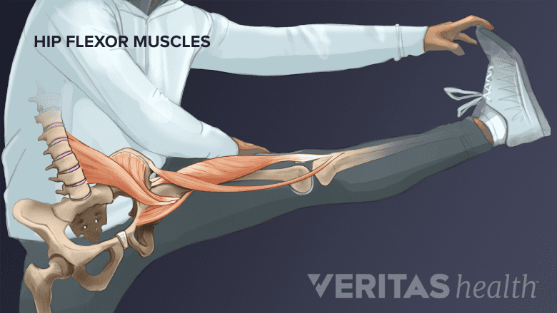
The flexor muscles of the hip help lift the leg and bend the knee.
The hip flexor muscles are situated at the front of the hip and are responsible for hip flexion – moving the leg forward or bringing the thigh toward the chest.
The primary hip flexors are:
- Iliopsoas
- Sartorius
- Tensor fasciae latae
- Rectus femoris
- Pectineus
- Adductor longus
The secondary hip flexors are:
- Adductor brevis
- Gracilis
- Gluteus minimus (anterior fibers)
The hip flexors are used while walking, running, cycling, and climbing stairs. For example, during walking, the iliopsoas contracts to lift the thigh forward with each step.
Hip Extensors
The hip extensor muscles are located at the back of the hip and buttock area and are responsible for hip extension – moving the leg backward.
The primary hip extensors are:
- Gluteus maximus
- Adductor magnus (posterior head)
- Biceps femoris (long head)
- Semitendinosus
- Semimembranosus
The secondary hip extensors are:
- Gluteus medius (middle and posterior fibers)
- Adductor magnus (anterior head)
The hip extensors are engaged in activities such as standing up from a seated position, climbing an uphill slope, ascending stairs, or pushing heavy objects. For example, when ascending stairs, the large gluteus maximus muscle in the buttock contracts to extend the hip as the leg moves backward to lift the body upward.
Hip Rotators
The hip’s rotator muscles lie deep within the hip joint and allow the thigh to turn inward (internal rotation) and outward (external rotation).
The primary hip rotators are:
- Gluteus maximus
- Piriformis
- Obturator internus
- Gemellus superior
- Gemellus inferior
- Quadratus femoris
The secondary hip rotators are:
- Gluteus medius (posterior fibers)
- Gluteus minimus (posterior fibers)
- Obturator externus
- Sartorius
- Biceps femoris (long head)
- Tensor fasciae latae
- Adductor longus
- Adductor brevis
- Pectineus
- Adductor magnus (posterior head)
The hip rotators are engaged in twisting movements in golf swings or ballet, or while taking a sharp turn while walking. For example, during a golf swing, the piriformis contracts to externally rotate the hip as the body twists to generate power in the swing.
Hip Adductors
The hip adductor muscles are positioned on the inner side of the thigh (groin) and bring the leg toward the body's midline (adduction).
The primary hip adductors are:
- Pectineus
- Adductor longus
- Gracilis
- Adductor brevis
- Adductor magnus (anterior and posterior heads)
The secondary hip adductors are:
- Biceps femoris (long head)
- Gluteus maximus (posterior fibers)
- Quadratus femoris
- Obturator externus
The hip adductors are involved in activities such as crossing the legs, bringing the legs together during swimming, or squeezing the legs together during hip thrusts.
For example, when performing swimming strokes such as the breaststroke, the hip adductors bring the legs together during the kicking motion. As the legs move outward and come together in a coordinated manner, the adductor muscles contract to pull the thighs toward the body's midline, generating propulsion and facilitating forward movement through the water.
Hip Abductors
The hip abductor muslces are located on the outer side of the hip and buttock and move the leg away from the body's midline.
The primary hip abductors are:
- Gluteus medius (all fibers)
- Gluteus minimus (all fibers)
- Tensor fasciae latae
The secondary hip abductors are:
- Piriformis
- Sartorius
- Rectus femoris
The abductors are engaged in activities such as side-stepping and balancing on one leg. For instance, during side-stepping exercises, the gluteus medius muscle on the outer side of the buttock contracts to stabilize the pelvis and maintain balance as the body moves sideways.
Hip Muscle Pain
The hip muscles can become weak or tight from prolonged sitting, insufficient warmups before participating in sports, and overexertion, causing pain and reduced mobility in the hip, buttock, and thigh.
Conditions that cause hip muscle and/or tendon pain include:
Strains and pulled muscles: Overstretching or tearing of the hip muscles, usually due to sudden movements or overuse, especially without adequate warm-up.
Tendinitis: Inflammation of the tendons around the hip, typically from repetitive stress or overuse
Myofascial pain syndrome: Chronic pain in the hip muscles due to trigger points, which are sensitive areas within the muscle fibers
Hip Ligament Anatomy
The hip ligaments are strong, fibrous bands of connective tissue that connect one bone to the other and reinforce the hip capsule (fibrous fluid-filled capsule surrounding the hip joint). They provide strength and stability to the joint and prevent excessive movement at the joint.
The major ligaments of the hip and their functions are described below2Glenister R, Sharma S. Anatomy, Bony Pelvis and Lower Limb, Hip. [Updated 2023 Jul 24]. In: StatPearls [Internet]. Treasure Island (FL): StatPearls Publishing; 2024 Jan-. Available from: https://www.ncbi.nlm.nih.gov/books/NBK526019/:
- Iliofemoral ligament (Y ligament of Bigelow): This triangle-shaped ligament is the strongest ligament in the body. It is located in the front part of the hip joint capsule and serves to:
- Prevent excessive hip joint extension (moving the leg backward) during activities like dancing or kicking.
- Maintain joint stability during activities like standing, walking, and running
- Pubofemoral ligament: This ligament is located in the front part of the hip joint capsule and serves to:
- Limit hip abduction (moving the leg away from the body’s midline)
- Limit hip extension (moving the leg backward)
- Maintain joint stability during movements such as standing up from a seated position or performing lateral movements like side-stepping
- Protect the joint from dislocation in an anterior (front) direct
- Ischiofemoral ligament: This ligament is located in the posterior (back) part of the hip joint capsule. It reinforces the posterior (back) part of the hip joint, limiting excessive internal rotation and adduction (moving the leg toward the body’s midline) of the hip. This stability is needed during activities such as squatting, climbing stairs, and bending forward at the hip.
- Zona orbicularis (annular ligament): This ligament surrounds the femoral head (upper part of the thighbone) within the acetabulum (hip socket) and forms a locking ring around the femoral head, securing it to the acetabulum. It serves to:
- Ensure joint stability during weight-bearing activities such as walking, running, and jumping.
- Facilitate smooth hip joint movement during hip rotation and flexion (bringing the knees toward the chest)
- Ligamentum teres: This pyramidal-shaped ligament connects the acetabulum to the femoral head from inside the joint. It serves to:
- Maintain joint stability by limiting excessive movement of the femoral head within the acetabulum
- Provide a pathway for blood vessels to nourish the femoral head in childhood and early adolescence
The iliofemoral, pubofemoral, and ischiofemoral ligaments together form the hip capsule.2
Hip Ligament Pain
The hip ligaments are susceptible to injury from trauma or overuse. Common ligament problems include:
- Ligament sprains: Over-stretching or tearing of the ligaments due to sudden movements, falls, or trauma. Hip sprains range from mild (micro-tears) to severe (complete tears).
- Capsular laxity: Overstretching the ligaments of the hip capsule due to overuse or trauma, leading to a feeling of instability.
Problems with the hip ligaments cause pain in and/or around the hip joint and reduced hip function.
Hip Bursae
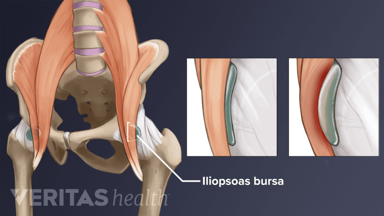
The iliopsoas bursa is located deep within the hip joint and reduces friction during hip movement.
Small, fluid-filled sacs called bursae are located near the hip joint and serve to reduce friction and cushion the movement of tendons and muscles over bones. Two main bursae in the hip region are:
- The trochanteric bursa. On the outer side of each hip, the trochanteric bursae cover the greater trochanter or prominent bony knob of the femur (thighbone). This bursa reduces friction between the iliotibial band (IT band) and the greater trochanter during hip movement.
- The iliopsoas bursa. Located deep within the hip joint, the iliopsoas bursa lies between the iliopsoas tendon and the underlying bone. It reduces friction between the iliopsoas tendon and the surrounding structures during hip movement.
The hip bursae tend to become inflamed and painful when there is repetitive friction, trauma to the hip region, or underlying hip conditions such as hip impingement or hip osteoarthritis. This condition is called hip bursitis.
Read about Hip Bursitis on Arthritis-health.com
- 1 Neumann DA. Kinesiology of the hip: a focus on muscular actions. J Orthop Sports Phys Ther. 2010;40(2):82-94. doi: 10.2519/jospt.2010.3025
- 2 Glenister R, Sharma S. Anatomy, Bony Pelvis and Lower Limb, Hip. [Updated 2023 Jul 24]. In: StatPearls [Internet]. Treasure Island (FL): StatPearls Publishing; 2024 Jan-. Available from: https://www.ncbi.nlm.nih.gov/books/NBK526019/